
- 阻害剤
- 研究分野別
- PI3K/Akt/mTOR
- Epigenetics
- Methylation
- Immunology & Inflammation
- Protein Tyrosine Kinase
- Angiogenesis
- Apoptosis
- Autophagy
- ER stress & UPR
- JAK/STAT
- MAPK
- Cytoskeletal Signaling
- Cell Cycle
- TGF-beta/Smad
- 化合物ライブラリー
- 抗体
- 新製品
- お問い合わせ
KU-55933
別名:ATM Kinase Inhibitor
KU-55933 is a potent and specific ATM inhibitor with IC50/Ki of 12.9 nM/2.2 nM in cell-free assays, and is highly selective for ATM as compared to DNA-PK, PI3K/PI4K, ATR and mTOR. KU‑55933 (ATM Kinase Inhibitor) inhibits the activation of autophagy‑initiating kinase ULK1 and results in a significant decrease of autophagy.
CAS No. 587871-26-9
文献中Selleckの製品使用例(396)
製品安全説明書
現在のバッチを見る:
純度:
99.95%
99.95
KU-55933関連製品
シグナル伝達経路
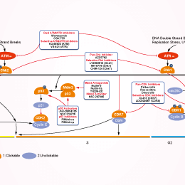
ATM/ATR阻害剤の選択性比較
Cell Data
| Cell Lines | Assay Type | Concentration | Incubation Time | 活性情報 | PMID |
|---|---|---|---|---|---|
| HepG2 | Growth Inhibition Assay | 10 μM | 24 h | blocks SC-III3-induced S phase arrest | 25527123 |
| HepG2 | Function Assay | 10 μM | 24 h | suppresses the phosphorylations of ATM on Ser1981, Chk1 on Ser345, Chk2 on Thr68, and Cdk2 on Tyr15 induced by SC-III3 | 25527123 |
| MCF10A | Growth Inhibition Assay | 10 μM | 24 h | potentiates the cytotoxicity of GA | 24150595 |
| HL-60 | Function Assay | 10 μM | 0.5 h | reduces phosphorylation of Chk2 | 23934411 |
| MCF-7 | Growth Inhibition Assay | 1-100μM | 24 h | inhibits the cell proliferation | 23185347 |
| HeLa | Growth Inhibition Assay | 1-100μM | 24 h | inhibits the cell proliferation | 23185347 |
| SH-SY5Y | Function Assay | 10 μM | 24 h | inhibits clioquinol-induced phosphorylation of p53 | 22627294 |
| IMR-32 | Function Assay | 10 μM | 24 h | inhibits clioquinol-induced phosphorylation of p53 | 22627294 |
| A549 | Function Assay | 10 μM | 1 h | suppresses Nano-Co-induced p53 accumulation | 22559321 |
| T47D | Function Assay | 20 mM | 24 h | prevents IR-induced degradation of IκBα | 21144805 |
| A29 MEF | Function Assay | 10 μM | 1h | blocks the phosphorylation of Akt at Ser473 | 20053781 |
| MDA-MB-453 | Growth Inhibition Assay | 5-40 μM | 72 h | IC50 of 10 μM | 20053781 |
| PC-3 | Growth Inhibition Assay | 5-40 μM | 72 h | IC50 of 10 μM | 20053781 |
| BJ | Function assay | 10 uM | 10 days | Suppression of senescence in human BJ cells assessed as increase in cell number at 10 uM after 10 days by senescence reversal assay | 16767085 |
| BJ | Function assay | 10 uM | 10 days | Inhibition of ataxia telangiectasia-mutated in human BJ cells assessed as increase in cell number at 10 uM after 10 days by senescence reversal assay | 16767085 |
| MCF7 | Function assay | 10 uM | 10 mins | Sensitization of infrared-induced DNA damage in human MCF7 cells assessed as reduction in colony formation at 10 uM pretreated for 10 mins followed by irradiation for 4 hrs measured after 10 days by crystal violet staining analysis | 26632965 |
| KATO III | Growth Inhibition Assay | 2.5/5/7.5 μM | enhances the toxicity of olaparib | 24841718 | |
| hTCEpi | Growth Inhibition Assay | 10 μM | prevents the cytopathic effect of HSV-1 | 24370835 | |
| MCF7 | Function assay | 1 hr | Inhibition of ATM kinase in human MCF7 cells after 1 hr by immunofluorescence assay, IC50 = 0.3 μM. | 26632965 | |
| KOSC-2 | Growth Inhibition Assay | IC50=26.9075 μM | SANGER | ||
| DEL | Growth Inhibition Assay | IC50=26.8356 μM | SANGER | ||
| GT3TKB | Growth Inhibition Assay | IC50=26.5342 μM | SANGER | ||
| MDA-MB-415 | Growth Inhibition Assay | IC50=26.5033 μM | SANGER | ||
| GI-1 | Growth Inhibition Assay | IC50=25.7055 μM | SANGER | ||
| BFTC-905 | Growth Inhibition Assay | IC50=25.5944 μM | SANGER | ||
| QIMR-WIL | Growth Inhibition Assay | IC50=25.1858 μM | SANGER | ||
| PANC-08-13 | Growth Inhibition Assay | IC50=25.0938 μM | SANGER | ||
| SK-MEL-30 | Growth Inhibition Assay | IC50=24.4662 μM | SANGER | ||
| CHL-1 | Growth Inhibition Assay | IC50=23.7292 μM | SANGER | ||
| Ramos-2G6-4C10 | Growth Inhibition Assay | IC50=22.96 μM | SANGER | ||
| SNU-449 | Growth Inhibition Assay | IC50=22.8748 μM | SANGER | ||
| HCC2157 | Growth Inhibition Assay | IC50=22.8054 μM | SANGER | ||
| LB1047-RCC | Growth Inhibition Assay | IC50=22.5879 μM | SANGER | ||
| YH-13 | Growth Inhibition Assay | IC50=22.5123 μM | SANGER | ||
| Mewo | Growth Inhibition Assay | IC50=22.5073 μM | SANGER | ||
| JVM-3 | Growth Inhibition Assay | IC50=22.506 μM | SANGER | ||
| HSC-3 | Growth Inhibition Assay | IC50=21.1835 μM | SANGER | ||
| U031 | Growth Inhibition Assay | IC50=21.1489 μM | SANGER | ||
| D-283MED | Growth Inhibition Assay | IC50=20.5339 μM | SANGER | ||
| A704 | Growth Inhibition Assay | IC50=19.8305 μM | SANGER | ||
| HCC70 | Growth Inhibition Assay | IC50=19.489 μM | SANGER | ||
| MLMA | Growth Inhibition Assay | IC50=19.0557 μM | SANGER | ||
| 697 | Growth Inhibition Assay | IC50=19.0201 μM | SANGER | ||
| HuP-T3 | Growth Inhibition Assay | IC50=18.5888 μM | SANGER | ||
| NCI-H2030 | Growth Inhibition Assay | IC50=18.1997 μM | SANGER | ||
| HCC2998 | Growth Inhibition Assay | IC50=17.6733 μM | SANGER | ||
| NCI-H82 | Growth Inhibition Assay | IC50=17.4573 μM | SANGER | ||
| CTB-1 | Growth Inhibition Assay | IC50=17.2259 μM | SANGER | ||
| NCI-SNU-1 | Growth Inhibition Assay | IC50=17.1269 μM | SANGER | ||
| SK-MEL-28 | Growth Inhibition Assay | IC50=17.0475 μM | SANGER | ||
| GCIY | Growth Inhibition Assay | IC50=16.7905 μM | SANGER | ||
| ChaGo-K-1 | Growth Inhibition Assay | IC50=16.6568 μM | SANGER | ||
| NCI-H1793 | Growth Inhibition Assay | IC50=16.4712 μM | SANGER | ||
| LXF-289 | Growth Inhibition Assay | IC50=16.2747 μM | SANGER | ||
| HuH-7 | Growth Inhibition Assay | IC50=16.2674 μM | SANGER | ||
| 8305C | Growth Inhibition Assay | IC50=16.1889 μM | SANGER | ||
| KG-1 | Growth Inhibition Assay | IC50=16.0996 μM | SANGER | ||
| VM-CUB-1 | Growth Inhibition Assay | IC50=15.9849 μM | SANGER | ||
| DBTRG-05MG | Growth Inhibition Assay | IC50=15.6111 μM | SANGER | ||
| D-423MG | Growth Inhibition Assay | IC50=15.5236 μM | SANGER | ||
| Hs-578-T | Growth Inhibition Assay | IC50=15.4182 μM | SANGER | ||
| DOK | Growth Inhibition Assay | IC50=15.3329 μM | SANGER | ||
| COLO-684 | Growth Inhibition Assay | IC50=14.1569 μM | SANGER | ||
| CAL-12T | Growth Inhibition Assay | IC50=13.617 μM | SANGER | ||
| KP-N-YS | Growth Inhibition Assay | IC50=12.6354 μM | SANGER | ||
| ES7 | Growth Inhibition Assay | IC50=11.788 μM | SANGER | ||
| LB2241-RCC | Growth Inhibition Assay | IC50=11.7186 μM | SANGER | ||
| GOTO | Growth Inhibition Assay | IC50=11.6996 μM | SANGER | ||
| J-RT3-T3-5 | Growth Inhibition Assay | IC50=11.2417 μM | SANGER | ||
| NCI-H1838 | Growth Inhibition Assay | IC50=11.1865 μM | SANGER | ||
| NCI-H1437 | Growth Inhibition Assay | IC50=9.8097 μM | SANGER | ||
| KM12 | Growth Inhibition Assay | IC50=9.21142 μM | SANGER | ||
| SK-MEL-3 | Growth Inhibition Assay | IC50=8.28575 μM | SANGER | ||
| HH | Growth Inhibition Assay | IC50=8.27671 μM | SANGER | ||
| LoVo | Growth Inhibition Assay | IC50=6.93239 μM | SANGER | ||
| CAL-72 | Growth Inhibition Assay | IC50=5.48084 μM | SANGER | ||
| LAMA-84 | Growth Inhibition Assay | IC50=4.58465 μM | SANGER | ||
| HuO-3N1 | Growth Inhibition Assay | IC50=4.17142 μM | SANGER | ||
| DU-145 | Growth Inhibition Assay | IC50=3.27352 μM | SANGER | ||
| RVH-421 | Growth Inhibition Assay | IC50=27.2921 μM | SANGER | ||
| EW-13 | Growth Inhibition Assay | IC50=27.4308 μM | SANGER | ||
| 639-V | Growth Inhibition Assay | IC50=27.5119 μM | SANGER | ||
| A2780 | Growth Inhibition Assay | IC50=27.641 μM | SANGER | ||
| SW982 | Growth Inhibition Assay | IC50=27.9052 μM | SANGER | ||
| SW1710 | Growth Inhibition Assay | IC50=28.0981 μM | SANGER | ||
| HCC1569 | Growth Inhibition Assay | IC50=28.4897 μM | SANGER | ||
| MV-4-11 | Growth Inhibition Assay | IC50=28.5735 μM | SANGER | ||
| BHT-101 | Growth Inhibition Assay | IC50=28.6572 μM | SANGER | ||
| Ca9-22 | Growth Inhibition Assay | IC50=28.714 μM | SANGER | ||
| HAL-01 | Growth Inhibition Assay | IC50=28.7615 μM | SANGER | ||
| D-263MG | Growth Inhibition Assay | IC50=29.344 μM | SANGER | ||
| NEC8 | Growth Inhibition Assay | IC50=29.5548 μM | SANGER | ||
| EKVX | Growth Inhibition Assay | IC50=31.5847 μM | SANGER | ||
| EM-2 | Growth Inhibition Assay | IC50=31.6304 μM | SANGER | ||
| MFM-223 | Growth Inhibition Assay | IC50=31.8098 μM | SANGER | ||
| SK-PN-DW | Growth Inhibition Assay | IC50=32.1406 μM | SANGER | ||
| HuO9 | Growth Inhibition Assay | IC50=32.5282 μM | SANGER | ||
| MHH-PREB-1 | Growth Inhibition Assay | IC50=32.6234 μM | SANGER | ||
| OVCAR-4 | Growth Inhibition Assay | IC50=32.8363 μM | SANGER | ||
| NCI-H1648 | Growth Inhibition Assay | IC50=32.8651 μM | SANGER | ||
| MKN1 | Growth Inhibition Assay | IC50=34.1101 μM | SANGER | ||
| KYSE-450 | Growth Inhibition Assay | IC50=34.6444 μM | SANGER | ||
| ES8 | Growth Inhibition Assay | IC50=34.8975 μM | SANGER | ||
| MS-1 | Growth Inhibition Assay | IC50=34.9554 μM | SANGER | ||
| HOP-92 | Growth Inhibition Assay | IC50=35.9277 μM | SANGER | ||
| SKG-IIIa | Growth Inhibition Assay | IC50=36.2561 μM | SANGER | ||
| TE-11 | Growth Inhibition Assay | IC50=36.5243 μM | SANGER | ||
| SK-NEP-1 | Growth Inhibition Assay | IC50=37.6744 μM | SANGER | ||
| DB | Growth Inhibition Assay | IC50=37.9185 μM | SANGER | ||
| IA-LM | Growth Inhibition Assay | IC50=38.0239 μM | SANGER | ||
| COLO-829 | Growth Inhibition Assay | IC50=38.4159 μM | SANGER | ||
| TGBC11TKB | Growth Inhibition Assay | IC50=39.1408 μM | SANGER | ||
| CAL-51 | Growth Inhibition Assay | IC50=40.0612 μM | SANGER | ||
| NCI-H2228 | Growth Inhibition Assay | IC50=40.3662 μM | SANGER | ||
| C32 | Growth Inhibition Assay | IC50=40.4024 μM | SANGER | ||
| KU-19-19 | Growth Inhibition Assay | IC50=40.7683 μM | SANGER | ||
| KNS-62 | Growth Inhibition Assay | IC50=40.8381 μM | SANGER | ||
| FADU | Growth Inhibition Assay | IC50=41.2502 μM | SANGER | ||
| CAL-33 | Growth Inhibition Assay | IC50=42.6749 μM | SANGER | ||
| CHP-134 | Growth Inhibition Assay | IC50=42.8496 μM | SANGER | ||
| HDLM-2 | Growth Inhibition Assay | IC50=42.9084 μM | SANGER | ||
| NBsusSR | Growth Inhibition Assay | IC50=43.0725 μM | SANGER | ||
| SW954 | Growth Inhibition Assay | IC50=43.1053 μM | SANGER | ||
| HCC1806 | Growth Inhibition Assay | IC50=43.411 μM | SANGER | ||
| VMRC-RCZ | Growth Inhibition Assay | IC50=43.4586 μM | SANGER | ||
| A549 | Growth Inhibition Assay | IC50=43.931 μM | SANGER | ||
| NKM-1 | Growth Inhibition Assay | IC50=43.9558 μM | SANGER | ||
| DMS-273 | Growth Inhibition Assay | IC50=44.7567 μM | SANGER | ||
| TYK-nu | Growth Inhibition Assay | IC50=45.1234 μM | SANGER | ||
| KALS-1 | Growth Inhibition Assay | IC50=45.146 μM | SANGER | ||
| A101D | Growth Inhibition Assay | IC50=45.4456 μM | SANGER | ||
| G-361 | Growth Inhibition Assay | IC50=46.2138 μM | SANGER | ||
| KARPAS-299 | Growth Inhibition Assay | IC50=46.3516 μM | SANGER | ||
| RS4-11 | Growth Inhibition Assay | IC50=46.542 μM | SANGER | ||
| HT-1376 | Growth Inhibition Assay | IC50=46.7426 μM | SANGER | ||
| SK-N-AS | Growth Inhibition Assay | IC50=46.7822 μM | SANGER | ||
| MG-63 | Growth Inhibition Assay | IC50=46.9036 μM | SANGER | ||
| EPLC-272H | Growth Inhibition Assay | IC50=46.9503 μM | SANGER | ||
| BALL-1 | Growth Inhibition Assay | IC50=47.832 μM | SANGER | ||
| LCLC-97TM1 | Growth Inhibition Assay | IC50=48.202 μM | SANGER | ||
| HO-1-N-1 | Growth Inhibition Assay | IC50=48.9676 μM | SANGER | ||
| MFE-280 | Growth Inhibition Assay | IC50=49.4617 μM | SANGER | ||
| NCI-H526 | Growth Inhibition Assay | IC50=49.8163 μM | SANGER | ||
| D-566MG | Growth Inhibition Assay | IC50=49.9096 μM | SANGER | ||
| BB30-HNC | Growth Inhibition Assay | IC50=49.9498 μM | SANGER | ||
| SK-N-DZ | Growth Inhibition Assay | IC50=50.0481 μM | SANGER | ||
| U2OS | Function assay | Inhibition of ATM in human U2OS cells assessed as inhibition of p53 phosphorylation at Ser15 residue, IC50 = 0.25 μM. | 26632965 | ||
| SK-N-MC | qHTS assay | qHTS of pediatric cancer cell lines to identify multiple opportunities for drug repurposing: Primary screen for SK-N-MC cells | 29435139 | ||
| 他の多くの細胞株試験データをご覧になる場合はこちらをクリックして下さい | |||||
生物活性
| 製品説明 | KU-55933 is a potent and specific ATM inhibitor with IC50/Ki of 12.9 nM/2.2 nM in cell-free assays, and is highly selective for ATM as compared to DNA-PK, PI3K/PI4K, ATR and mTOR. KU‑55933 (ATM Kinase Inhibitor) inhibits the activation of autophagy‑initiating kinase ULK1 and results in a significant decrease of autophagy. | ||
|---|---|---|---|
| Targets |
|
| In Vitro | ||||
| In vitro | KU-55933 inhibits DNA-PK and PI3K with IC50 of 2.5 μM and 16.6 μM, respectively. Besides, this compound also prevents the activity of mTOR with IC50 of 9.3 μM. It is active at the cellular level in ablating a well-characterized ATM-dependent phosphorylation event. This chemical has a dose-dependent effect in inhibiting this ATM-dependent phosphorylation event with IC50 of 300 nM. KU-58050 does not prevent the ATM-dependent phosphorylation of p53 serine 15 until a dose of 30 μM. Addition of this compound has no appreciable effects on UV-induced phosphorylation of H2AX on serine 139, NBS1 on serine 343, CHK1 on serine 345, and SMC1 on serine 966. In stark contrast to the UV responses, it ablates the ionizing radiation-induced phosphorylation of these ATM substrates. This chemical sensitizes HeLa cells to a range of ionizing radiation doses. [1] It inhibits the phosphorylation of Akt induced by growth factors in cancer cells. This compound suppresses the proliferation of cancer cells. Furthermore, suppression of ATM by this chemical improves survival, probably via prevention of downstream activation of TAp63α. [2] | |||
|---|---|---|---|---|
| Kinase Assay | Purified enzyme assays | |||
| ATM for use in the in vitro assay is obtained from HeLa nuclear extract by immunoprecipitation with rabbit polyclonal antiserum raised to the COOH-terminal 400 amino acids of ATM in buffer containing 25 mM HEPES (pH 7.4), 2 mM MgCl2, 250 mM KCl, 500 μM EDTA, 100 μM Na3VO4, 10% v/v glycerol, and 0.1% v/v Igepal. ATM-antibody complexes are isolated from nuclear extract by incubating with protein A-Sepharose beads for 1 hour and then through centrifugation to recover the beads. In the well of a 96-well plate, ATM-containing Sepharose beads are incubated with 1 μg of substrate glutathione S-transferase–p53N66 (NH2-terminal 66 amino acids of p53 fused to glutathione S-transferase) in ATM assay buffer [25 mM HEPES (pH 7.4), 75 mM NaCl, 3 mM MgCl2, 2 mM MnCl2, 50 μM Na3VO4, 500 μM DTT, and 5% v/v glycerol] at 37 °C in the presence or absence of this compound. After 10 minutes with gentle shaking, ATP is added to a final concentration of 50 μM and the reaction continued at 37 °C for an additional 1 hour. The plate is centrifuged at 250 × g for 10 minutes (4 °C) to remove the ATM-containing beads, and the supernatant is removed and transferred to a white opaque 96-well plate and incubated at room temperature for 1.5 hours to allow glutathione S-transferase-p53N66 binding. This plate is then washed with PBS, blotted dry, and analyzed by a standard ELISA technique with a phospho-serine 15 p53 antibody. The detection of phosphorylated glutathione S-transferase-p53N66 substrate is performed in combination with a goat antimouse horseradish peroxidase-conjugated secondary antibody. Enhanced chemiluminescence solution is used to produce a signal and chemiluminescent detection is carried out. | ||||
| 細胞実験 | 細胞株 | U2OS cells | ||
| 濃度 | 10 μM | |||
| 反応時間 | 2 hours | |||
| 実験の流れ | U2OS cells are exposed to ionizing radiation (3, 5, or 15 Gy) or UV (5 or 50 J/m2) and the ATM response determined by Western blot analysis of p53 serine 15 phosphorylation and stabilization of wild-type p53. Whole cell extracts are obtained from each time point, proteins separated by SDS-PAGE, and the ATM-specific increase in phosphorylated serine 15 measured with a p53 phospho-serine 15 specific antibody. Overall p53 stabilization with time is also observed with a p53-specific antibody (DO-1). Similarly, for studying ATM-dependent phosphorylations on H2AX, CHK1, NBS1, and SMC1, the following antibodies are used: CHK1 phospho-serine 345 and NBS1 phospho-serine 343 antibodies. Histone H2A (H-124) and CHK1 antibodies are also used, as well as SMC1 and SMC1 phospho-serine 966 antibodies. For determination of a cellular IC50 for KU-55933, the peak response time for p53 serine 15 phosphorylation of 2 hours is used to monitor inhibition of ATM. This compound is titrated onto cells and preincubated for 1 hour before ionizing radiation. Using scanning densitometry, the percentage inhibition relative to vehicle control is calculated, and the IC50 value is calculated as for the in vitro determinations. |
|||
| 実験結果図 | Methods | Biomarkers | 結果図 | PMID |
| Western blot | p-AKT(Ser473) / p-AKT(Thr308) PARP / Cleaved PARP / Caspase-3 / Cleaved caspase-3 ATM-S1981 / ATM / p-p53(S15) / p53 p21 / p27 / p53 |
|
22739265 | |
| Immunofluorescence | p-S824 KAP1 / ZEBRA |
|
28249048 | |
| In Vivo | ||
| In Vivo | Suppression of ATM-dependent STAT3 activation by KU-55933 enhances TRAIL-mediated apoptosis through up-regulation of surface DR5 expression, whereas suppression of both STAT3 and NF-κB appeares to be involved in down-regulation of cFLIP accompanied by an additional increase in apoptotic levels. This compound affects TRAIL-mediated apoptosis more strongly than the JAK2 inhibitor, AG490, or overexpression of STAT3β. [3] | |
|---|---|---|
| 動物実験 | 動物モデル | BALB/c nu/nu nude mice bearing LU1205 cells |
| 投与量 | 10 μM | |
| 投与経路 | -- | |
|
化学情報
| 分子量 | 395.49 | 化学式 | C21H17NO3S2 |
| CAS No. | 587871-26-9 | SDF | Download KU-55933 SDFをダウンロードする |
| Smiles | C1COCCN1C2=CC(=O)C=C(O2)C3=C4C(=CC=C3)SC5=CC=CC=C5S4 | ||
| 保管 | |||
|
In vitro |
DMSO : 39 mg/mL ( (98.61 mM); 吸湿したDMSOは溶解度を減少させます。新しいDMSOをご使用ください。) Water : Insoluble Ethanol : Insoluble |
モル濃度計算器 |
|
in vivo Add solvents to the product individually and in order. |
投与溶液組成計算機 | |||||
実験計算
投与溶液組成計算機(クリア溶液)
ステップ1:実験データを入力してください。(実験操作によるロスを考慮し、動物数を1匹分多くして計算・調製することを推奨します)
mg/kg
g
μL
匹
ステップ2:投与溶媒の組成を入力してください。(ロット毎に適した溶解組成が異なる場合があります。詳細については弊社までお問い合わせください)
% DMSO
%
% Tween 80
% ddH2O
%DMSO
%
計算結果:
投与溶媒濃度: mg/ml;
DMSOストック溶液調製方法: mg 試薬を μL DMSOに溶解する(濃度 mg/mL, 注:濃度が当該ロットのDMSO溶解度を超える場合はご連絡ください。 )
投与溶媒調製方法:Take μL DMSOストック溶液に μL PEG300,を加え、完全溶解後μL Tween 80,を加えて完全溶解させた後 μL ddH2O,を加え完全に溶解させます。
投与溶媒調製方法:μL DMSOストック溶液に μL Corn oil,を加え、完全溶解。
注意:1.ストック溶液に沈殿、混濁などがないことをご確認ください;
2.順番通りに溶剤を加えてください。次のステップに進む前に溶液に沈殿、混濁などがないことを確認してから加えてください。ボルテックス、ソニケーション、水浴加熱など物理的な方法で溶解を早めることは可能です。
技術サポート
ストックの作り方、阻害剤の保管方法、細胞実験や動物実験の際に注意すべき点など、製品を取扱う時に問い合わせが多かった質問に対しては取扱説明書でお答えしています。
他に質問がある場合は、お気軽にお問い合わせください。
* 必須
納期 国内在庫品:受注日の翌日(15時までの受注分) *北海道、九州、沖縄への配送は受注日より2日以上 を要する場合あり 海外在庫品:受注後1〜2週間