
- 阻害剤
- 研究分野別
- PI3K/Akt/mTOR
- Epigenetics
- Methylation
- Immunology & Inflammation
- Protein Tyrosine Kinase
- Angiogenesis
- Apoptosis
- Autophagy
- ER stress & UPR
- JAK/STAT
- MAPK
- Cytoskeletal Signaling
- Cell Cycle
- TGF-beta/Smad
- 化合物ライブラリー
- 抗体
- 新製品
- お問い合わせ
GSK1070916
GSK1070916 is a reversible and ATP-competitive inhibitor of Aurora B/C with IC50 of 3.5 nM/6.5 nM. It displays >100-fold selectivity against the closely related Aurora A-TPX2 complex. Phase 1.
CAS No. 942918-07-2
文献中Selleckの製品使用例(12)
カスタマーフィードバック4例
製品安全説明書
現在のバッチを見る:
S274003
DMSO]
93 mg/mL]
false]
Ethanol]
8 mg/mL]
false]
Water]
Insoluble]
false
純度:
99.65%
99.65
GSK1070916関連製品
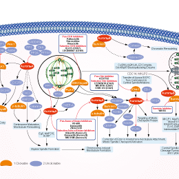
シグナル伝達経路
Aurora Kinase阻害剤の選択性比較
Cell Data
| Cell Lines | Assay Type | Concentration | Incubation Time | 活性情報 | PMID |
|---|---|---|---|---|---|
| A549 cells | Proliferation assay | 6-7 d | Antiproliferative activity against human A549 cells after 6 to 7 days by celltiter-glo luminescence assay in absence of 70% human serum albumin, EC50=0.007 μM | 20420387 | |
| 他の多くの細胞株試験データをご覧になる場合はこちらをクリックして下さい | |||||
生物活性
| 製品説明 | GSK1070916 is a reversible and ATP-competitive inhibitor of Aurora B/C with IC50 of 3.5 nM/6.5 nM. It displays >100-fold selectivity against the closely related Aurora A-TPX2 complex. Phase 1. | |||||||||||
|---|---|---|---|---|---|---|---|---|---|---|---|---|
| Targets |
|
| In Vitro | ||||
| In vitro | GSK1070916 selectively inhibits Aurora B and Aurora C with Ki of 0.38 nM and 1.5 nM over Aurora A with Ki of 490 nM. Inhibition of Aurora B and Aurora C is time-dependent, with an enzyme-inhibitor dissociation half-life of >480 min and 270 min respectively. In addition, GSK1070916 is also a competitive inhibitor with respect to ATP. [1] Human tumor cells treated with GSK1070916 shows dose-dependent inhibition of phosphorylation on serine 10 of Histone H3, a substrate specific for Aurora B. Moreover, GSK1070916 inhibits the proliferation of tumor cells with EC50 values of <10 nM in over 100 cell lines spanning a broad range of tumor types, with a median EC50 of 8 nM. Although GSK1070916 has potent activity against proliferating cells, a dramatic shift in potency is observed in primary, nondividing, normal human vein endothelial cells. Furthermore, GSK1070916-treated cells do not arrest in mitosis but instead fails to divide and become polyploid, ultimately leading to apoptosis. [2] In another study, it is also reported high chromosome number associated with resistance to the inhibition of Aurora B and C suggests cells with a mechanism to bypass the high ploidy checkpoint are resistant to GSK1070916. [3] | |||
|---|---|---|---|---|
| Kinase Assay | Kinase Assay | |||
| The ability of GSK1070916 to inhibit the Aurora enzymes is measured using in vivo kinase assays. The assays measure the ability of Aurora A, Aurora B and Aurora C to phosphorylate a synthetic peptide substrate. Biotin-Ahx-RARRRLSFFFFAKKK-NH2 is used for the Aurora A–TPX2 LEADseekerTM assay and 5FAM-PKAtide is used for the IMAPTM assay for all three Aurora kinases. To take into account time-dependent inhibition of Aurora enzymes, Aurora A–TPX2, Aurora B–INCENP and Aurora C–INCENP are incubated with GSK1070916 at various concentrations for 30 min before the reactions are initiated with the addition of substrates. For the Aurora A LEADseekerTM assay, final assay conditions are 0.5 nM Aurora A–TPX2, 1 μM peptide substrate, 6 mM MgCl2, 1.5 μM ATP, 0.003 μCi/μL [γ-33P] ATP in 50 mM Hepes, pH 7.2, 0.15 mg/mL BSA, 0.01% Tween-20, 5 mM DTT and 25 mM KCl. The reactions are incubated at room temperature (25 °C) for 120 min and terminated by the addition of LEADseekerTM beads in PBS containing EDTA (final concentration 2 mg/mL beads and 25 mM EDTA). The plates are then sealed, and the beads are allowed to settle overnight. Product formation is quantified using a Viewlux Imager. For the IMAPTM assays, Aurora A–TPX2 (final concentration 1 nM), Aurora B–INCENP (final concentration 2 nM) or Aurora C–INCENP (final concentration 2.5 nM) is added to the compound-containing plates in 5 μL of buffer (25 mM Hepes, pH 7.2, for Aurora A, 25 mM Hepes, pH 7.5, for Aurora B and 20 mM Hepes, pH 7.2, for Aurora C) containing 0.15 mg/mL BSA, 0.01% Tween 20 and 25 mM NaCl. This mixture is incubated at room temperature for 30 min. To start the reaction, 5 μL of a substrate solution is added containing the same Hepes buffer as used for the pre-incubation, 25 mM NaCl, MgCl2 (2, 4 and 4 mM for Aurora A, B and C respectively), DTT (4, 4 and 2 mM for Aurora A, B and C respectively), ATP (4, 4 and 10 μM for Aurora A, B and C respectively), 200 nM 5FAM-PKAtide, 0.01% Tween 20 and 0.15 mg/mL BSA. The reactions are incubated at room temperature for 120 min for Aurora A and B and 60 min for Aurora C. These reactions are then terminated by the addition of 10 μL of 1:500 (1:600 for Aurora C) Progressive Binding Reagent in 95% Progressive Binding Buffer A and 5% Progressive Binding Buffer B. Plates are incubated at room temperature for approx. 90–120 min (time allowed for equilibrium to be reached). Plates are read in a Molecular Devices Analyst plate reader in fluorescence polarization mode. | ||||
| 細胞実験 | 細胞株 | SW48, Colo 201, SW480, WiDr, Colo205, RKO E6, RKO, LoVo, HCT-116, SW620, HT29, W1417, DLD-1, HCT-8, Colo 320HSR, Hep-3B, OVCAR-3, MEC-1 cells | ||
| 濃度 | 0-15 mM | |||
| 反応時間 | 6-7 days | |||
| 実験の流れ | Cells are plated in 96-well plates in the recommended growth media and incubated at 37 °C in 5% CO2 overnight. The following day, the cells are treated with serial dilutions of GSK1070916. At this time, one set of cells is treated with CellTiter-Glo for a time equal to 0 (T = 0) measurement. Following a 6- to 7-d incubation with compound, cell proliferation is measured using the CellTiter-Glo reagent according to the manufacture's recommended protocol. As inhibition of Aurora B induces endomitosis, the degree of which differs depending on the cell type, an extended compound treatment time is required to accurately reflect the effects on cell viability across a large panel of cell lines. For analysis of cell viability, values from wells with no cells are subtracted for background correction and the data plotted as a percent of the DMSO-treated control samples using Microsoft Excel XLfit4 software. The EC50 values represent the concentration of GSK1070916 where 50% maximal effect is observed | |||
| In Vivo | ||
| In Vivo | GSK1070916 (25, 50, or 100 mg/kg) shows dose-dependent inhibition of phosphorylation of an Aurora B–specific substrate in mice and consistent with its broad cellular activity, has antitumor effects in 10 human tumor xenograft models including breast, colon, lung, and two leukemia models. [2] | |
|---|---|---|
| 動物実験 | 動物モデル | Mice tumor xenograft models (A549, SW620, HCT116, H460, MCF-7, HL60, K562, Colo205) |
| 投与量 | 25, 50, or 100 mg/kg | |
| 投与経路 | Administered via i.p. once daily | |
化学情報
| 分子量 | 507.63 | 化学式 | C30H33N7O |
| CAS No. | 942918-07-2 | SDF | Download GSK1070916 SDFをダウンロードする |
| Smiles | CCN1C=C(C(=N1)C2=CC=C(C=C2)NC(=O)N(C)C)C3=C4C=C(NC4=NC=C3)C5=CC=CC(=C5)CN(C)C | ||
| 保管 | |||
|
In vitro |
DMSO : 93 mg/mL ( (183.2 mM); 吸湿したDMSOは溶解度を減少させます。新しいDMSOをご使用ください。) Ethanol : 8 mg/mL Water : Insoluble |
モル濃度計算器 |
|
in vivo Add solvents to the product individually and in order. |
投与溶液組成計算機 | ||||
実験計算
投与溶液組成計算機(クリア溶液)
ステップ1:実験データを入力してください。(実験操作によるロスを考慮し、動物数を1匹分多くして計算・調製することを推奨します)
mg/kg
g
μL
匹
ステップ2:投与溶媒の組成を入力してください。(ロット毎に適した溶解組成が異なる場合があります。詳細については弊社までお問い合わせください)
% DMSO
%
% Tween 80
% ddH2O
%DMSO
%
計算結果:
投与溶媒濃度: mg/ml;
DMSOストック溶液調製方法: mg 試薬を μL DMSOに溶解する(濃度 mg/mL, 注:濃度が当該ロットのDMSO溶解度を超える場合はご連絡ください。 )
投与溶媒調製方法:Take μL DMSOストック溶液に μL PEG300,を加え、完全溶解後μL Tween 80,を加えて完全溶解させた後 μL ddH2O,を加え完全に溶解させます。
投与溶媒調製方法:μL DMSOストック溶液に μL Corn oil,を加え、完全溶解。
注意:1.ストック溶液に沈殿、混濁などがないことをご確認ください;
2.順番通りに溶剤を加えてください。次のステップに進む前に溶液に沈殿、混濁などがないことを確認してから加えてください。ボルテックス、ソニケーション、水浴加熱など物理的な方法で溶解を早めることは可能です。
技術サポート
ストックの作り方、阻害剤の保管方法、細胞実験や動物実験の際に注意すべき点など、製品を取扱う時に問い合わせが多かった質問に対しては取扱説明書でお答えしています。
他に質問がある場合は、お気軽にお問い合わせください。
* 必須
Tags: GSK1070916を買う | GSK1070916 ic50 | GSK1070916供給者 | GSK1070916を購入する | GSK1070916費用 | GSK1070916生産者 | オーダーGSK1070916 | GSK1070916化学構造 | GSK1070916分子量 | GSK1070916代理店
納期 国内在庫品:受注日の翌日(15時までの受注分) *北海道、九州、沖縄への配送は受注日より2日以上 を要する場合あり 海外在庫品:受注後1〜2週間