- 阻害剤
- 研究分野別
- PI3K/Akt/mTOR
- Epigenetics
- Methylation
- Immunology & Inflammation
- Protein Tyrosine Kinase
- Angiogenesis
- Apoptosis
- Autophagy
- ER stress & UPR
- JAK/STAT
- MAPK
- Cytoskeletal Signaling
- Cell Cycle
- TGF-beta/Smad
- 化合物ライブラリー
- Popular Compound Libraries
- Customize Library
- Clinical and FDA-approved Related
- Bioactive Compound Libraries
- Inhibitor Related
- Natural Product Related
- Metabolism Related
- Cell Death Related
- By Signaling Pathway
- By Disease
- Anti-infection and Antiviral Related
- Neuronal and Immunology Related
- Fragment and Covalent Related
- FDA-approved Drug Library
- FDA-approved & Passed Phase I Drug Library
- Preclinical/Clinical Compound Library
- Bioactive Compound Library-I
- Bioactive Compound Library-II
- Kinase Inhibitor Library
- Express-Pick Library
- Natural Product Library
- Human Endogenous Metabolite Compound Library
- Alkaloid Compound LibraryNew
- Angiogenesis Related compound Library
- Anti-Aging Compound Library
- Anti-alzheimer Disease Compound Library
- Antibiotics compound Library
- Anti-cancer Compound Library
- Anti-cancer Compound Library-Ⅱ
- Anti-cancer Metabolism Compound Library
- Anti-Cardiovascular Disease Compound Library
- Anti-diabetic Compound Library
- Anti-infection Compound Library
- Antioxidant Compound Library
- Anti-parasitic Compound Library
- Antiviral Compound Library
- Apoptosis Compound Library
- Autophagy Compound Library
- Calcium Channel Blocker LibraryNew
- Cambridge Cancer Compound Library
- Carbohydrate Metabolism Compound LibraryNew
- Cell Cycle compound library
- CNS-Penetrant Compound Library
- Covalent Inhibitor Library
- Cytokine Inhibitor LibraryNew
- Cytoskeletal Signaling Pathway Compound Library
- DNA Damage/DNA Repair compound Library
- Drug-like Compound Library
- Endoplasmic Reticulum Stress Compound Library
- Epigenetics Compound Library
- Exosome Secretion Related Compound LibraryNew
- FDA-approved Anticancer Drug LibraryNew
- Ferroptosis Compound Library
- Flavonoid Compound Library
- Fragment Library
- Glutamine Metabolism Compound Library
- Glycolysis Compound Library
- GPCR Compound Library
- Gut Microbial Metabolite Library
- HIF-1 Signaling Pathway Compound Library
- Highly Selective Inhibitor Library
- Histone modification compound library
- HTS Library for Drug Discovery
- Human Hormone Related Compound LibraryNew
- Human Transcription Factor Compound LibraryNew
- Immunology/Inflammation Compound Library
- Inhibitor Library
- Ion Channel Ligand Library
- JAK/STAT compound library
- Lipid Metabolism Compound LibraryNew
- Macrocyclic Compound Library
- MAPK Inhibitor Library
- Medicine Food Homology Compound Library
- Metabolism Compound Library
- Methylation Compound Library
- Mouse Metabolite Compound LibraryNew
- Natural Organic Compound Library
- Neuronal Signaling Compound Library
- NF-κB Signaling Compound Library
- Nucleoside Analogue Library
- Obesity Compound Library
- Oxidative Stress Compound LibraryNew
- Phenotypic Screening Library
- PI3K/Akt Inhibitor Library
- Protease Inhibitor Library
- Protein-protein Interaction Inhibitor Library
- Pyroptosis Compound Library
- Small Molecule Immuno-Oncology Compound Library
- Mitochondria-Targeted Compound LibraryNew
- Stem Cell Differentiation Compound LibraryNew
- Stem Cell Signaling Compound Library
- Natural Phenol Compound LibraryNew
- Natural Terpenoid Compound LibraryNew
- TGF-beta/Smad compound library
- Traditional Chinese Medicine Library
- Tyrosine Kinase Inhibitor Library
- Ubiquitination Compound Library
-
Cherry Picking
You can personalize your library with chemicals from within Selleck's inventory. Build the right library for your research endeavors by choosing from compounds in all of our available libraries.
Please contact us at info@selleck.co.jp to customize your library.
You could select:
- 抗体
- 新製品
- お問い合わせ
PP2
別名:AG 1879,AGL 1879
PP2 (AG 1879, AGL 1879), a Src family kinase inhibitor, potently inhibits Lck/Fyn with IC50 of 4 nM/5 nM in cell-free assays, ~100-fold less potent to EGFR, inactive for ZAP-70, JAK2 and PKA.

CAS No. 172889-27-9
文献中Selleckの製品使用例(127)
製品安全説明書
現在のバッチを見る:
純度:
99.5%
99.5
PP2関連製品
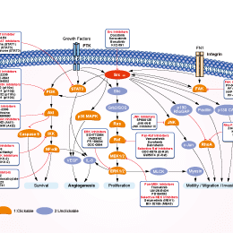
シグナル伝達経路
Src阻害剤の選択性比較
Cell Data
| Cell Lines | Assay Type | Concentration | Incubation Time | 活性情報 | PMID |
|---|---|---|---|---|---|
| A431 | Function assay | 10 uM | Inhibition of Src autophosphorylation of Y419 in A431 cells at 10 uM | 16509573 | |
| 8701-BC | Proapoptotic assay | 10 uM | Proapoptotic activity against 8701-BC cells at 10 uM by PARP assay | 16509573 | |
| Saos2 | Cytotoxicity assay | 48 hrs | Cytotoxicity against human Saos2 cells after 48 hrs by MTT assay, IC50 = 8.07 μM. | 23932070 | |
| SH-SY5Y | Antiproliferative assay | 72 hrs | Antiproliferative activity against human SH-SY5Y cells assessed as cell viability after 72 hrs by XTT assay, IC50 = 6.1 μM. | 21856155 | |
| SaOS2 | Antiproliferative assay | Antiproliferative activity against human SaOS2 cells assessed as cellular viability, IC50 = 8.1 μM. | 17929792 | ||
| T-cells | Function assay | Inhibition of tyrosine phosphorylation in human T cells, IC50 = 0.6 μM. | 18077363 | ||
| T-cells | Function assay | Inhibition of adhesion kinase in human T cells, IC50 = 0.6 μM. | 18077363 | ||
| A549 | Growth inhibition assay | Growth inhibition of human A549 cells, IC50 = 0.01 μM. | 28814374 | ||
| A431 | Function assay | Inhibitory effect on phospho-Src/nonphospho after EGF (100 uM) stimulation of A431 cells (21), IC50 = 17 μM. | 15109642 | ||
| MEG01 | Antiproliferative assay | Antiproliferative activity against human MEG01 cells, IC50 = 17 μM. | 18257513 | ||
| A431 | Function assay | Inhibitory effect on phospho-Src (Tyr416) after EGF (100 uM) stimulation of A431 cells (38), IC50 = 22 μM. | 15109642 | ||
| K562 | Antiproliferative assay | Antiproliferative activity against human K562 cells, IC50 = 25 μM. | 18257513 | ||
| A431 | Antiproliferative assay | Tested for antiproliferative activity against human A431 cells, IC50 = 32 μM. | 15109642 | ||
| A431 | Antiproliferative assay | Antiproliferative activity against A431 cells, IC50 = 32.2 μM. | 16509573 | ||
| KU812 | Antiproliferative assay | Antiproliferative activity against human KU812 cells, IC50 = 45 μM. | 18257513 | ||
| 他の多くの細胞株試験データをご覧になる場合はこちらをクリックして下さい | |||||
生物活性
| 製品説明 | PP2 (AG 1879, AGL 1879), a Src family kinase inhibitor, potently inhibits Lck/Fyn with IC50 of 4 nM/5 nM in cell-free assays, ~100-fold less potent to EGFR, inactive for ZAP-70, JAK2 and PKA. | ||||
|---|---|---|---|---|---|
| Targets |
|
| In Vitro | ||||
| In vitro |
PP2 inhibits Src by binding to an area of the molecule that does not overlap with the ATP binding domain. [2] This compound (20 μM) induces 40-50% growth inhibition of HT29 cells, this concentration reduces the Src activity as early as 1 hour and maintains a 35% inhibition of Src activity for 2 days. It (100 mM) decreases the Src activity of HT29 cells in a dose-dependent manner. This chemical (1 mM-100 mM) causes a dose-dependent growth inhibition of human colon cancer cells (HT29, SW480, and PMCO1), liver cancer cells (PLC/PRF/5, KYN-2, Li7, and HepG2), and breast cancer cells (MCF-7, MDA-MB-468, and BT-474). It (20 μM) significantly increases aggregation in most of the cancer cells (HT29, SW480, PMCO1, PLC/PRF/5, KYN-2, Li7, MCF-7, and MDA-MB-468) in E-cadherin dependent manner. This compound (20 μM) enhances E-cadherin expression and also strongly increases E-cadherin’s association with the actin cytoskeleton in cancer cells. It (20 μM) increases the expression of α-catenin, β-catenin, and γ-catenin in HT29 cells, whereas in PLC/PRF/5 and MCF-7 cells, the total protein level of α-catenin does not change, but the levels of β- catenin and γ-catenin increases slightly. [3] This inhibitor inhibits proliferation of two cervical cancer cells (HeLa and SiHa) in a time- and dose-dependent manner. It (10 μM) down-regulates pSrc-Y416, pEGFR-Y845, and -Y1173 expression levels in HeLa and SiHa cells. This chemical (10 μM) could modulate cell cycle arrest by up-regulating p21(Cip1) and p27(Kip1) in both HeLa and SiHa cells and down-regulating expression of cyclin A, and cyclin dependent kinase-2, -4 (Cdk-2, -4) in HeLa and of cyclin B and Cdk-2 in SiHa. [4] |
|||
|---|---|---|---|---|
| Kinase Assay | Immune complex enzyme assays | |||
| The acid-treated enolase is diluted 1:20 with 1× PBS before aliquoting 100 mL/well into a Nunc 96-well high protein binding assay plate. Assay wells are then aspirated; blocked with 0.5% bovine serum, 1× PBS for 1 h at 37 ℃;and then washed five times with 300 mL of 1× PBS/well. The source of Lck is either LSTRA cells or Lck expressed in HeLa cells using a vaccinia expression system. FynT is expressed in HeLa cells using the vaccinia system. Cells (12.5×10⁶/mL) are lysed in lysis buffer, and the lysates are clarified by centrifugation at 14,000 cpm for 15 min at 4 ℃ in an Eppendorf tube. The clarified lysates are then incubated with the appropriate anti-kinase antibody at 10 μg/mL for 2 h at 4 ℃. Protein A-Sepharose beads are added to the antibody/lysate mixture at 250 μL/mL and allowed to incubate for 30 min at 4 ℃. The beads are then washed twice in 1 mL of lysis buffer and twice in 1 mL of kinase buffer (25 mM HEPES, 3 mM MnCl2, 5mMMgCl2, and 100 μM sodium orthovanadate) and resuspended to 50% (w/v) in kinase buffer. Twenty-five microliters of the bead suspension is added to each well of the enolase-coated 96-well high protein binding plate together with an appropriate concentration of this compound and [γ-32P]ATP (25 μL/well of a 200 μCi/mL solution in kinase buffer). After incubation for 20 min at 20 ℃, 60 μLl of boiling 2× solubilization buffer containing 10 mM ATP is added to the assay wells to terminate the reactions. Thirty microliters of the samples is removed from the wells, boiled for 5 min, and run on a 7.5% SDS-polyacrylamide gel. The gels are subsequently dried and exposed to Kodak X-AR film. For quantitation, films are scanned using a Molecular Dynamics laser scanner, and the optical density of the major substrate band, enolase p46, is determined. In companion experiments for measuring the activity of this chemical against Lck, the assay plate is washed with two wash cycles on a Skatron harvester using 50 mM EDTA, 1 mM ATP. Scintillation fluid (100 μL) is then added to the wells, and 32P incorporation is measured using a micro-β-counter. | ||||
| 細胞実験 | 細胞株 | HT29, SW480, PMCO1, PLC/PRF/5, KYN-2, Li7, HepG2, MCF-7, MDA-MB-468 and BT-474 cell lines | ||
| 濃度 | ~100 μM | |||
| 反応時間 | 2 days | |||
| 実験の流れ | Cell viability is determined using an in vitro toxicology assay kit following the manufacturer’s instructions. Cells are seeded in 96-well plates at day 0. Starting at day 1, cells are treated for 2 days with each of a series of increasing concentrations of PP2 (1 μM, 10 μM, and 100 μM). At the end of this period, cell proliferation is evaluated by mitochondria dehydrogenase in viable cells, leading to formazan formation. This experiment is repeated three times with 10 determinations/tested concentration. |
|||
| 実験結果図 | Methods | Biomarkers | 結果図 | PMID |
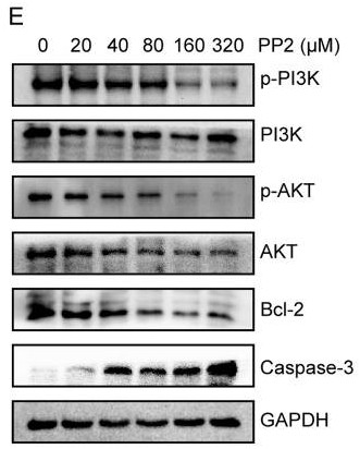
| Western blot | p-PI3K / PI3K / p-AKT / AKT / Bcl-2 / Caspase-3 p-Src / Src / p-MAPK / MAPK |

|
30250573 | |
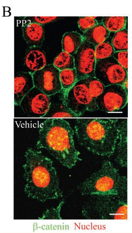
| Immunofluorescence | β-catenin FAK / p-FAK |

|
18566211 | |
| In Vivo | ||
| In Vivo |
PP2 (5 mg/kg/day) induces some slowing in the growth rate of the primary tumors relative to the control treated with vehicle in SCID mice inoculated HT29 cells in the spleen. This compound induces some slowing in the growth rate of the primary tumors relative to the control treated with vehicle in SCID mice inoculated HT29 cells in the spleen. This chemical significantly reduces the relative liver weight and liver metastasis volume compared with the controls in SCID mice inoculated HT29 cells in the spleen. [3] This compound (1.5 mg/kg i.p.) treated rats show approximately 50% reduction of infarct size on T2-weighted MRI and in TTC staining compared with controls in rats with focal ischemic brain injury. This chemical results in better the neurological score than controls in rats with focal ischemic brain injury. [5] |
|
|---|---|---|
| 動物実験 | 動物モデル | SCID mice inoculated HT29 cells in the spleen |
| 投与量 | 5 mg/kg/day | |
| 投与経路 | intraperitoneal injection | |
| NCT Number | Recruitment | Conditions | Sponsor/Collaborators | Start Date | Phases |
|---|---|---|---|---|---|
| NCT03842371 | Unknown status | Sepsis Syndrome |
West China Hospital |
February 11 2019 | -- |
| NCT02407626 | Terminated | Myocardial Ischemia |
Triemli Hospital|University of Alberta |
September 2015 | Not Applicable |
| NCT02315287 | Unknown status | Type 2 Diabetes |
Seoul National University Bundang Hospital |
September 2014 | Phase 4 |
|
化学情報
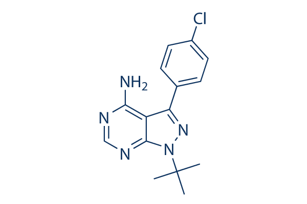
| 分子量 | 301.77 | 化学式 | C15H16ClN5 |
| CAS No. | 172889-27-9 | SDF | Download PP2 SDFをダウンロードする |
| Smiles | CC(C)(C)N1C2=NC=NC(=C2C(=N1)C3=CC=C(C=C3)Cl)N | ||
| 保管 | |||
|
In vitro |
DMSO : 60 mg/mL ( (198.82 mM); 吸湿したDMSOは溶解度を減少させます。新しいDMSOをご使用ください。) Ethanol : 2 mg/mL Water : Insoluble |
モル濃度計算器 |
|
in vivo Add solvents to the product individually and in order. |
投与溶液組成計算機 | |||||
実験計算
投与溶液組成計算機(クリア溶液)
ステップ1:実験データを入力してください。(実験操作によるロスを考慮し、動物数を1匹分多くして計算・調製することを推奨します)
mg/kg
g
μL
匹
ステップ2:投与溶媒の組成を入力してください。(ロット毎に適した溶解組成が異なる場合があります。詳細については弊社までお問い合わせください)
% DMSO
%
% Tween 80
% ddH2O
%DMSO
%
計算結果:
投与溶媒濃度: mg/ml;
DMSOストック溶液調製方法: mg 試薬を μL DMSOに溶解する(濃度 mg/mL, 注:濃度が当該ロットのDMSO溶解度を超える場合はご連絡ください。 )
投与溶媒調製方法:Take μL DMSOストック溶液に μL PEG300,を加え、完全溶解後μL Tween 80,を加えて完全溶解させた後 μL ddH2O,を加え完全に溶解させます。
投与溶媒調製方法:μL DMSOストック溶液に μL Corn oil,を加え、完全溶解。
注意:1.ストック溶液に沈殿、混濁などがないことをご確認ください;
2.順番通りに溶剤を加えてください。次のステップに進む前に溶液に沈殿、混濁などがないことを確認してから加えてください。ボルテックス、ソニケーション、水浴加熱など物理的な方法で溶解を早めることは可能です。
技術サポート
ストックの作り方、阻害剤の保管方法、細胞実験や動物実験の際に注意すべき点など、製品を取扱う時に問い合わせが多かった質問に対しては取扱説明書でお答えしています。
他に質問がある場合は、お気軽にお問い合わせください。
* 必須
よくある質問(FAQ)
質問1:
Could you please suggest me the in vivo details about the dilution to reduce the amount of DMSO to 1 to 5% for it?
回答
For in vivo study, we recommend to use 4% DMSO +Corn oil up to 2.5 mg/ml for it.

