
- 阻害剤
- 研究分野別
- PI3K/Akt/mTOR
- Epigenetics
- Methylation
- Immunology & Inflammation
- Protein Tyrosine Kinase
- Angiogenesis
- Apoptosis
- Autophagy
- ER stress & UPR
- JAK/STAT
- MAPK
- Cytoskeletal Signaling
- Cell Cycle
- TGF-beta/Smad
- 化合物ライブラリー
- 抗体
- 新製品
- お問い合わせ
Glucose Transporter GLUT1 Rabbit mAb
Catalog No.: F1192
-
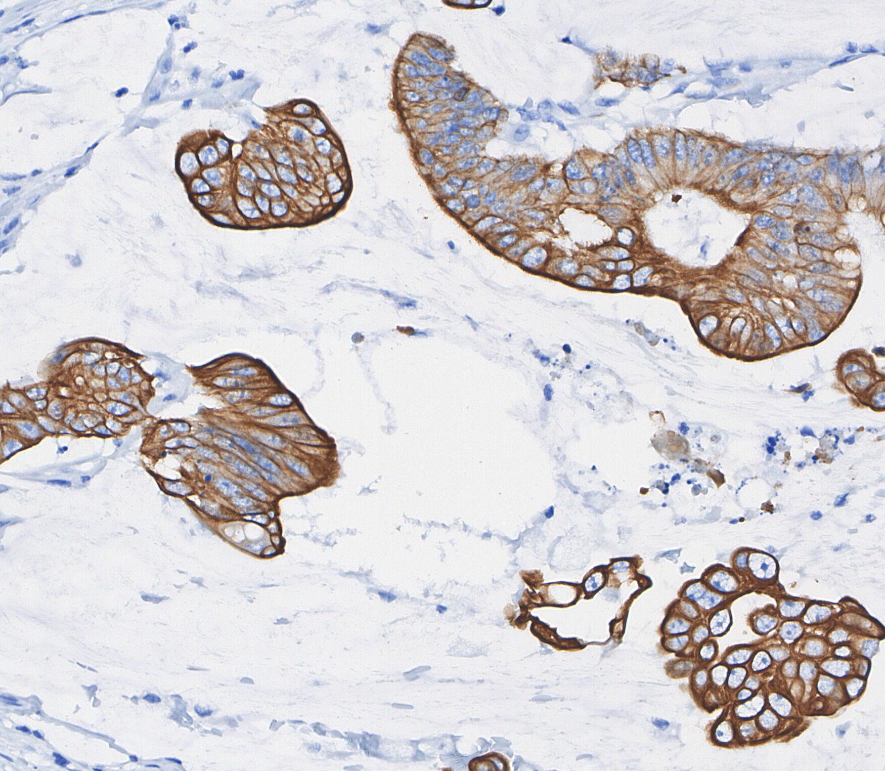 Immunohistochemical analysis of formalin fixed paraffin embedded human Colorectal cancer tissue with F1192 at 1/100 dilution.
カスタマーフィードバック(2)
キーポイント
タンパク質の局在:細胞膜 (PMID:18245775)。
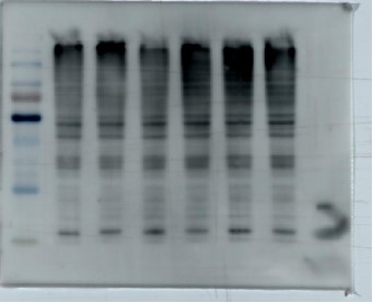
WB
RIPA/NP-40 Lysis Buffer バッファーでのライセート調製を推奨します。
推奨一次抗体の希釈倍率 1:10000。
WB
RIPA/NP-40 Lysis Buffer バッファーでのライセート調製を推奨します。
推奨一次抗体の希釈倍率 1:10000。
よくある問題とその対策
使用情報
| Dilution |
|---|
|
| Application |
|---|
| WB, IHC, IF, FCM |
| Source |
|---|
| Rabbit |
| Reactivity |
|---|
| Human, Mouse, Rat |
| Storage Buffer |
|---|
| PBS, pH 7.2+50% Glycerol+0.05% BSA+0.01% NaN₃ |
| Storage (from the date of receipt) |
|---|
| –20°C (avoid freeze-thaw cycles), 2 years |
| Predicted MW |
|---|
| 54 kDa |
| ポジティブコントロール | Human placenta; human fetal liver; Human colonic adenocarcinoma; Human cervical carcinoma; Human lung carcinoma; Mouse liver; Mouse brain; Human fetal brain; Human normal skin; Human normal breast; Human normal colon; Human kidney carcinoma; Human skeletal muscle; Human urinary bladder transitional carcinoma; HT-29; 3T3-L1; SW480; Jurkat; HeLa; HepG2; A549; Jurkat |
|---|---|
| ネガティブコントロール |
サンプル処理データの例
| サンプル | 処理状況 |
| PDAC | Transfection (WDR79)(PMID:36712589) |
| HepG2 | DMSO (20 μM, 24 h) (PMID:36048765) |
| NCI-N87 | Ca2+(2 μM, 37 ℃, 10h)(PMID:38693181) |
| クリックして、さらに多くのサンプルデータを表示 | |
*異なるヒト由来細胞や組織における発現量の予測については、以下をご参照ください: http://www.proteinatlas.org
プロトコール
| WB |
|---|
Experimental Protocol:
Sample preparation
1. Tissue: Lyse the tissue sample by adding an appropriate volume of ice-cold RIPA/NP-40 Lysis Buffer (containing Protease Inhibitor Cocktail),and homogenize the tissue at a low temperature.
2. Adherent cell: Aspirate the culture medium and wash the cells with ice-cold PBS twice. Lyse the cells by adding an appropriate volume of RIPA/NP-40 Lysis Buffer (containing Protease Inhibitor Cocktail) and put the sample on ice for 5 min.
3. Suspension cell: Transfer the culture medium to a pre-cooled centrifuge tube. Centrifuge and aspirate the supernatant. Wash the cells with ice-cold PBS twice. Lyse the cells by adding an appropriate volume of RIPA/NP-40 Lysis Buffer (containing Protease Inhibitor Cocktail) and put the sample on ice for 5 min.
4. Place the lysate into a pre-cooled microcentrifuge tube. Centrifuge at 4°C for 15 min. Collect the supernatant;
5. Remove a small volume of lysate to determine the protein concentration;
6. Add protein loading buffer to the 20 μL sample, and keep it on ice for immediate use; or heat the sample at 50~70 ℃, then let it cool on ice and centrifuge for 5 min.
Electrophoretic separation
1. According to the concentration of extracted protein, load appropriate amount of protein sample and marker onto SDS-PAGE gels for electrophoresis. Recommended separating gel (lower gel) concentration: 10%. Reference Table for Selecting SDS-PAGE Separation Gel Concentrations
2. Power up 80V for 30 minutes. Then the power supply is adjusted (110 V~150 V), the Marker is observed, and the electrophoresis can be stopped when the indicator band of the predyed protein Marker where the protein is located is properly separated. (Note that the current should not be too large when electrophoresis, too large current (more than 150 mA) will cause the temperature to rise, affecting the result of running glue. If high currents cannot be avoided, an ice bath can be used to cool the bath.)
Transfer membrane
1. Take out the converter, soak the clip and consumables in the pre-cooled converter;
2. Activate PVDF membrane with methanol for 1 min and rinse with transfer buffer;
3. Install it in the order of "black edge of clip - sponge - filter paper - filter paper - glue -PVDF membrane - filter paper - filter paper - sponge - white edge of clip";
4. The protein was electrotransferred to PVDF membrane. ( 0.45 µm PVDF membrane is recommended ) Reference Table for Selecting PVDF Membrane Pore Size Specifications
Recommended conditions for wet transfer: 200 mA, 120 min.
( Note that the transfer conditions can be adjusted according to the protein size. For high-molecular-weight proteins, a higher current and longer transfer time are recommended. However, ensure that the transfer tank remains at a low temperature to prevent gel melting.)
Block
1. After electrotransfer, wash the film with TBST at room temperature for 5 minutes;
2. Incubate the film in the blocking solution for 1 hour at room temperature;
3. Wash the film with TBST for 3 times, 5 minutes each time.
Antibody incubation
1. Use 5% skim milk powder to prepare the primary antibody working liquid (recommended dilution ratio for primary antibody 1:10000), gently shake and incubate with the film at 4°C overnight;
2. Wash the film with TBST 3 times, 5 minutes each time;
3. Add the secondary antibody to the blocking solution and incubate with the film gently at room temperature for 1 hour;
4. After incubation, wash the film with TBST 3 times for 5 minutes each time.
Antibody staining
1. Add the prepared ECL luminescent substrate (or select other color developing substrate according to the second antibody) and mix evenly;
2. Incubate with the film for 1 minute, remove excess substrate (keep the film moist), wrap with plastic film, and expose in the imaging system.
|
| IF |
|---|
Experimental Protocol:
Sample Preparation
1. Adherent Cells: Place a clean, sterile coverslip in a culture dish. Once the cells grow to near confluence as a monolayer, remove the coverslip for further use.
2. Suspension Cells: Seed the cells onto a clean, sterile slide coated with poly-L-lysine.
3. Frozen Sections: Allow the slide to thaw at room temperature. Wash it with pure water or PBS for 2 times, 3 minutes each time.
4. Paraffin Sections: Deparaffinization and rehydration. Wash the slide with pure water or PBS for 3 times, 3 minutes each time. Then perform antigen retrieval.
Fixation
1. Fix the cell coverslips/spots or tissue sections at room temperature using a fixative such as 4% paraformaldehyde (4% PFA) for 10-15 minutes.
2. Wash the sample with PBS for 3 times, 3 minutes each time.
Blocking
Add blocking solution and incubate at room temperature for at least 1 hour. (Common blocking solutions include: serum from the same source as the secondary antibody, BSA, or goat serum.)
Note: Ensure the sample remains moist during and after the blocking step to prevent drying, which can lead to high background.
Immunofluorescence Staining (Day 1)
1. Remove the blocking solution and add the diluted primary antibody.
2. Incubate the sample in a humidified chamber at 4°C overnight.
Immunofluorescence Staining (Day 2)
1. Remove the primary antibody and wash with PBST for 3 times, 5 minutes each time.
2. Add the diluted fluorescent secondary antibody and incubate in the dark at 4°C for 1–2 hours.
3. Remove the secondary antibody and wash with PBST for 3 times, 5 minutes each time.
4. Add diluted DAPI and incubate at room temperature in the dark for 5–10 minutes.
5. Wash with PBST for 3 times, 5 minutes each time.
Mounting
1. Mount the sample with an anti-fade mounting medium.
2. Allow the slide to dry at room temperature overnight in the dark.
3. Store the slide in a slide storage box at 4°C, protected from light.
|
生物学的記述
| Specificity |
|---|
Glucose Transporter GLUT1 Rabbit mAb detects endogenous levels of total Glucose Transporter GLUT1 protein. |
| Synonym(s) |
|---|
| Glucose Transporter GLUT1,Glut1 |
| Uniprot ID |
|---|
| P11166 |
| Clone |
|---|
| P22E15 |
| Background |
|---|
GLUTs, or facilitated diffusion glucose transporters, are membrane proteins with 12 membrane-spanning regions and intracellular amino and carboxyl terminals. They facilitate glucose transport across the plasma membrane through a facilitated diffusion mechanism. Based on sequence identity and alignment studies, GLUTs are divided into three subclasses: Class I, II, and III. Class I includes GLUT1 to GLUT4, with GLUT1 being predominantly expressed in human beta cells. In hepatocytes, GLUT1 is involved in bi-directional glucose transport regulated by hormones like thyroid hormone. |
| References |
|---|
技術サポート
ストックの作り方、阻害剤の保管方法、細胞実験や動物実験の際に注意すべき点など、製品を取扱う時に問い合わせが多かった質問に対しては取扱説明書でお答えしています。
他に質問がある場合は、お気軽にお問い合わせください。
* 必須
納期 国内在庫品:受注日の翌日(15時までの受注分) *北海道、九州、沖縄への配送は受注日より2日以上 を要する場合あり 海外在庫品:受注後1〜2週間