
- 阻害剤
- 研究分野別
- PI3K/Akt/mTOR
- Epigenetics
- Methylation
- Immunology & Inflammation
- Protein Tyrosine Kinase
- Angiogenesis
- Apoptosis
- Autophagy
- ER stress & UPR
- JAK/STAT
- MAPK
- Cytoskeletal Signaling
- Cell Cycle
- TGF-beta/Smad
- 化合物ライブラリー
- FDA-approved Drug Library
- FDA-approved & Passed Phase I Drug Library
- Preclinical/Clinical Compound Library
- Bioactive Compound Library-I
- Bioactive Compound Library-II
- Kinase Inhibitor Library
- Express-Pick Library
- Natural Product Library
- Human Endogenous Metabolite Compound Library
- Covalent Inhibitor Library
- FDA-approved Anticancer Drug LibraryNew
- Highly Selective Inhibitor Library
- HTS Library for Drug Discovery
- Metabolism Compound Library
- 抗体
- 新製品
- お問い合わせ
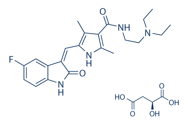
Sunitinib malate
別名:SU11248 malate
Sunitinib malate is a multi-targeted RTK inhibitor targeting VEGFR2 (Flk-1) and PDGFRβ with IC50 of 80 nM and 2 nM in cell-free assays, and also inhibits c-Kit. Sunitinib Malate effectively inhibits autophosphorylation of Ire1α. Sunitinib Malate increases both death receptor and mitochondrial-dependent apoptosis.
CAS No. 341031-54-7
文献中Selleckの製品使用例(166)
製品安全説明書
Sunitinib malate関連製品
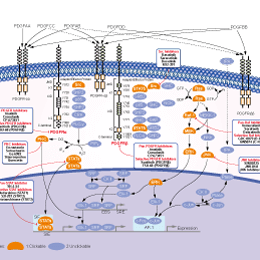
シグナル伝達経路
PDGFR阻害剤の選択性比較
Cell Data
| Cell Lines | Assay Type | Concentration | Incubation Time | 活性情報 | PMID |
|---|---|---|---|---|---|
| U251 | Kinase Assay | 10 μM | 1 h | Inhibition of VEGFR2 with IC50 of 0.0189 μM | 24890652 |
| SH-SY5Y | Kinase Assay | 10 μM | 1 h | Inhibition of PDGFRbeta with IC50 of 0.0831 μM | 24890652 |
| A431 | Kinase Assay | 10 μM | 1 h | Inhibition of EGFR with IC50 of 0.1721 μM | 24890652 |
| 他の多くの細胞株試験データをご覧になる場合はこちらをクリックして下さい | |||||
生物活性
| 製品説明 | Sunitinib malate is a multi-targeted RTK inhibitor targeting VEGFR2 (Flk-1) and PDGFRβ with IC50 of 80 nM and 2 nM in cell-free assays, and also inhibits c-Kit. Sunitinib Malate effectively inhibits autophosphorylation of Ire1α. Sunitinib Malate increases both death receptor and mitochondrial-dependent apoptosis. | ||||||||||
|---|---|---|---|---|---|---|---|---|---|---|---|
| Targets |
|
| In Vitro | ||||
| In vitro |
Sunitinib also potently inhibits Kit and FLT-3. [1] Sunitinib is a potent ATP-competitive inhibitor of VEGFR2 (Flk1) and PDGFRβ with Ki of 9 nM and 8 nM, respectively, displaying >10-fold higher selectivity for VEGFR2 and PDGFR than FGFR-1, EGFR, Cdk2, Met, IGFR-1, Abl, and src. In serum-starved NIH-3T3 cells expressing VEGFR2 or PDGFRβ, Sunitinib inhibits VEGF-dependent VEGFR2 phosphorylation and PDGF-dependent PDGFRβ phosphorylation with IC50 of 10 nM and 10 nM, respectively. Sunitinib inhibits VEGF-induced proliferation of serum-starved HUVECs with IC50 of 40 nM, and inhibits PDGF-induced proliferation of NIH-3T3 cells overexpressing PDGFRβ or PDGFRα with IC50 of 39 nM and 69 nM, respectively. [2] Sunitinib inhibits phosphorylation of wild-type FLT3, FLT3-ITD, and FLT3-Asp835 with IC50 of 250 nM, 50 nM, and 30 nM, respectively. Sunitinib inhibits the proliferation of MV4;11 and OC1-AML5 cells with IC50 of 8 nM and 14 nM, respectively, and induces apoptosis in a dose-dependent manner. [3] |
|||
|---|---|---|---|---|
| Kinase Assay | Biochemical Tyrosine Kinase Assays | |||
| IC50 values for Sunitinib against VEGFR2 (Flk-1) and PDGFRβ are determined using glutathione S-transferasefusion proteins containing the complete cytoplasmic domain of the RTK. Biochemical tyrosine kinase assays to quantitate the trans-phosphorylation activity of VEGFR2 (Flk-1) and PDGFRβ are performed in 96-well microtiter plates precoated (20 μg/well in PBS; incubated overnight at 4 °C) with the peptide substrate poly-Glu,Tyr (4:1). Excess protein binding sites are blocked with the addition of 1-5% (w/v) BSA in PBS. Purified GST-fusion proteins are produced in baculovirus-infected insect cells. GST-VEGFR2 and GST-PDGFRβ are then added to the microtiter wells in 2 × concentration kinase dilution buffer consisting of 100 mM HEPES, 50 mM NaCl, 40 μM NaVO4, and 0.02% (w/v) BSA. The final enzyme concentration for GST-VEGFR2 or GST-PDGFRβ is 50 ng/mL. Twenty-five μL of diluted Sunitinib are subsequently added to each reaction well to produce a range of inhibitor concentrations appropriate for each enzyme. The kinase reaction is initiated by the addition of different concentrations of ATP in a solution of MnCl2 so that the final ATP concentrations spanned the Km for the enzyme, and the final concentration of MnCl2 is 10 mM. The plates are incubated for 5-15 minutes at room temperature before stopping the reaction with the addition of EDTA. The plates are then washed three times with TBST. Rabbit polyclonal antiphosphotyrosine antisera are added to the wells at a 1:10,000 dilution in TBST containing 0.5% (w/v) BSA, 0.025% (w/v) nonfat dry milk, and 100 μM NaVO4 and incubated for 1 hour at 37 °C. The plates are then washed three times with TBST, followed by the addition of goat antirabbit antisera conjugated with horseradish peroxidase (1:10,000 dilution in TBST). The plates are incubated for 1 hour at 37 °C and then washed three times with TBST. The amount of phosphotyrosine in each well is quantitated after the addition of 2,2′-azino-di-[3-ethylbenzthiazoline sulfonate] as substrate. | ||||
| 細胞実験 | 細胞株 | RS4;11, MV4;11, and OC1-AML5 | ||
| 濃度 | Dissolved in DMSO, final concentrations ~10 μM | |||
| 反応時間 | 24 and 48 hours | |||
| 実験の流れ | Cells are starved overnight in medium containing 0.1% FBS prior to addition of Sunitinib and FL (50 ng/mL; FLT3-WT cells only). Proliferation is measured after 48 hours of culture using the Alamar Blue assay or trypan blue cell viability assays. Apoptosis is measured 24 hours after Sunitinib addition by Western blotting to detect cleavage of poly (ADP-ribose) polymerase (PARP) or levels of caspase-3. |
|||
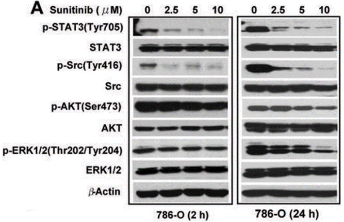
| 実験結果図 | Methods | Biomarkers | 結果図 | PMID |
| Western blot | p-STAT3 / STAT3 / p-Src / Src / p-AKT / AKT / p-ERK / ERK p-GSK3β / GSK3β / MYCN p-AKT / AKT / p-ERK / ERK |
|
19244102 | |
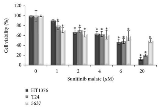
| Growth inhibition assay | Cell viability |
|
24369536 | |
| In Vivo | ||
| In Vivo |
Consistent with the substantial and selective inhibition of VEGFR2 or PDGFR phosphorylation and signaling in vivo, Sunitinib (20-80 mg/kg/day) exhibits broad and potent dose-dependent anti-tumor activity against a variety of tumor xenograft models including HT-29, A431, Colo205, H-460, SF763T, C6, A375, or MDA-MB-435. Sunitinib dosing at 80 mg/kg/day for 21 days leads to complete tumor regression in six of eight mice, without tumor re-growing during a 110-day observation period after the end of treatment. Second round of treatment with Sunitinib remains efficacious against tumors that are not fully regressed during the first round of treatment. Sunitinib treatment results in significant decrease in tumor MVD, with ~40% reduction in SF763T glioma tumors. SU11248 treatment results in a complete inhibition of additional tumor growth of luciferase-expressing PC-3M xenografts, despite no reduction in tumor size. [2] Sunitinib treatment (20 mg/kg/day) dramatically suppresses the growth subcutaneous MV4;11 (FLT3-ITD) xenografts and prolongs survival in the FLT3-ITD bone marrow engraftment model. [3] |
|
|---|---|---|
| 動物実験 | 動物モデル | Female nu/nu mice implanted s.c. with HT-29, A431, Colo205, H-460, SF763T, C6, A375, or MDA-MB-435, and male nu/nu mice bearing luciferase-expressing PC-3M tumors |
| 投与量 | ~80 mg/kg | |
| 投与経路 | Orally once daily | |
| NCT Number | Recruitment | Conditions | Sponsor/Collaborators | Start Date | Phases |
|---|---|---|---|---|---|
| NCT06222593 | Not yet recruiting | Carcinoma Renal Cell |
State University of New York at Buffalo |
June 1 2024 | Phase 1|Phase 2 |
| NCT06208748 | Not yet recruiting | Gastrointestinal Stromal Tumors|GIST |
Sarcoma Alliance for Research through Collaboration|Cogent Biosciences Inc.|Dana-Farber Cancer Institute|The Life Raft Group |
March 2024 | Phase 2 |
| NCT05745142 | Completed | Carcinoma Renal Cell|Clear-cell Metastatic Renal Cell Carcinoma |
Pfizer |
February 23 2023 | -- |
| NCT05043090 | Recruiting | Papillary Renal Cell Carcinoma |
AstraZeneca |
October 28 2021 | Phase 3 |
| NCT04669366 | Completed | Kidney Neoplasms |
Pfizer |
January 20 2021 | -- |
化学情報
| 分子量 | 532.56 | 化学式 | C22H27FN4O2.C4H6O5 |
| CAS No. | 341031-54-7 | SDF | Download Sunitinib malate SDFをダウンロードする |
| Smiles | CCN(CC)CCNC(=O)C1=C(NC(=C1C)C=C2C3=C(C=CC(=C3)F)NC2=O)C.C(C(C(=O)O)O)C(=O)O | ||
| 保管 | |||
|
In vitro |
DMSO : 70 mg/mL ( (131.44 mM); 吸湿したDMSOは溶解度を減少させます。新しいDMSOをご使用ください。) Water : Insoluble Ethanol : Insoluble |
モル濃度計算器 |
|
in vivo Add solvents to the product individually and in order. |
投与溶液組成計算機 | ||||
| Clear solution |
5%DMSO
Corn oil
|
5.0mg/ml (9.39mM) | Taking the 1 mL working solution as an example, add 50 μL of 100 mg/ml clear DMSO stock solution to 950 μL of corn oil and mix evenly. The mixed solution should be used immediately for optimal results. | ||
実験計算
投与溶液組成計算機(クリア溶液)
ステップ1:実験データを入力してください。(実験操作によるロスを考慮し、動物数を1匹分多くして計算・調製することを推奨します)
mg/kg
g
μL
匹
ステップ2:投与溶媒の組成を入力してください。(ロット毎に適した溶解組成が異なる場合があります。詳細については弊社までお問い合わせください)
% DMSO
%
% Tween 80
% ddH2O
%DMSO
%
計算結果:
投与溶媒濃度: mg/ml;
DMSOストック溶液調製方法: mg 試薬を μL DMSOに溶解する(濃度 mg/mL, 注:濃度が当該ロットのDMSO溶解度を超える場合はご連絡ください。 )
投与溶媒調製方法:Take μL DMSOストック溶液に μL PEG300,を加え、完全溶解後μL Tween 80,を加えて完全溶解させた後 μL ddH2O,を加え完全に溶解させます。
投与溶媒調製方法:μL DMSOストック溶液に μL Corn oil,を加え、完全溶解。
注意:1.ストック溶液に沈殿、混濁などがないことをご確認ください;
2.順番通りに溶剤を加えてください。次のステップに進む前に溶液に沈殿、混濁などがないことを確認してから加えてください。ボルテックス、ソニケーション、水浴加熱など物理的な方法で溶解を早めることは可能です。
技術サポート
ストックの作り方、阻害剤の保管方法、細胞実験や動物実験の際に注意すべき点など、製品を取扱う時に問い合わせが多かった質問に対しては取扱説明書でお答えしています。
他に質問がある場合は、お気軽にお問い合わせください。
* 必須
よくある質問(FAQ)
質問1:
I was wondering that the compound is in its cis or trans form?
回答
S1042 Sunitinib Malate is Z form.
質問2:
What is the difference between Sunitinib Malate(S1042) and Sunitinib(S7781)?
回答
S1042 is the Malate salt form of Sunitinib. The biological activities of these two compounds are the same but the solubility of these two compounds in aqueous solvent are different.
Tags: Sunitinib malateを買う | Sunitinib malate ic50 | Sunitinib malate供給者 | Sunitinib malateを購入する | Sunitinib malate費用 | Sunitinib malate生産者 | オーダーSunitinib malate | Sunitinib malate化学構造 | Sunitinib malate分子量 | Sunitinib malate代理店
納期
国内在庫品:受注日の翌日(15時までの受注分)
*北海道、九州、沖縄への配送は受注日より2日以上
を要する場合あり
海外在庫品:受注後1〜2週間